DLS Sizing of Adeno-Associated Viruses (AAVs)
Joelle Medinger, Coline Bretz
Related Product
LS Spectrometer™ II
The LS SpectrometerTM II is a goniometer-based variable-angle light scattering instrument for static- (SLS) and dynamic light scattering (DLS). The LS SpectrometerTM II allows for the most comprehensive nanoparticle characterization and can be further upgraded with various options, such as Modulated 3D technology for the measurement of turbid samples.
Introduction
The adeno-associated virus (AAV) is a prominent vector used in gene therapy, thanks to its low immune response and wide host-cell range. Among the critical quality attributes (CQAs) necessary to control for proper manufacturing, aggregation has a particular influence on the stability and shelf-life of therapeutic formulations[1]. Analytical techniques such AUC, TEM, or SEC are used to characterize such formulations and measure possible aggregates. However, these techniques are time-consuming, destructive, and incur low throughput[2]. In this context, Dynamic Light Scattering (DLS), when carried out using advanced instrumentation, provides a straightforward and rapid way of evaluating the particle size distribution (PSD) in AAV formulations and detecting the presence of aggregates - responsible for product degradation - with high sensitivity and in-situ. In this application report, we present DLS sizing results performed on a popular AAV serotype, using a highly powerful DLS instrument.
Materials & Methods
An AAV9 sample containing no DNA payload and labeled “empty” was received from Virovek Inc (USA). No dilution or filtration was performed. The sample was measured on the goniometer-based variable multi-angle light scattering instrument (V-MALS) 3D LS SpectrometerTM from LS Instruments equipped with a 100 mW laser with a 660 nm wavelength. The measurement angle was set to 90°, and the measurement was carried out with activated modulated 3D technology to remove any possible artifacts from multiple scattering[4]. 10 repetitions of 60 seconds were carried out. The results were obtained by means of the CORENN algorithm[3].
Results & Discussion
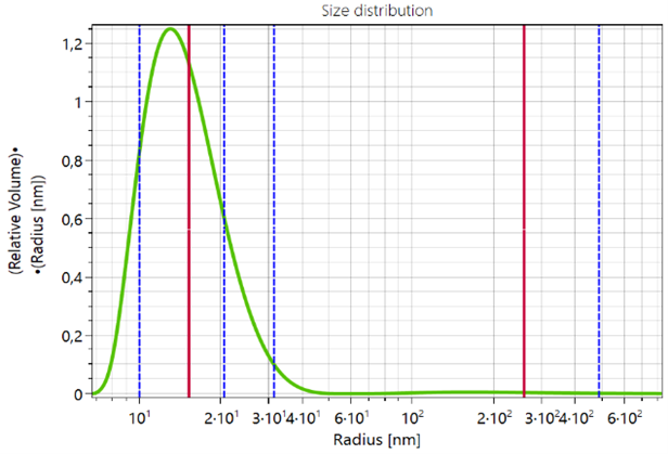
Fig1 shows a representative PSD measured for the AAV9 “empty” sample.

Fig. 1: Volume-weighted particle size distribution of the AAV9 “empty” sample.
A main particle population with an average (red vertical bar) diameter of 30 nm is identified. We also notice the presence of a small amount of aggregates with an average size of 310 nm. The averaged results are summarized in the following table:
|
Population |
Amount [%] |
Radius [nm] |
PDI |
|
1 |
99 |
14.5 |
0.81 |
|
2 |
1 |
310 |
0.49 |
Conclusion
Using a popular AAV serotype, we show that such systems can be characterized in a fast and straightforward manner using advanced DLS. The sensitivity provided by novel fitting algorithms and high laser power enables proper detection of low amounts of aggregates.
Authors
Joelle Medinger, Coline Bretz
References
[1] Wright, J. F. et al., Gene Therapy, 2008, 15, 840–848.
[2] Gimpel, A. et al., Mol. Ther. - Methods Clin. Dev., 2021, 20, 740–754.
[3] https://lsinstruments.ch/en/theory/dynamic-light-scattering-dls/dls-data-analysis-the-corenn-method.
[4] https://lsinstruments.ch/en/theory/dynamic-light-scattering-dls/modulated-3d-cross-correlation.
