Characterizing Concentrated Colloidal Suspensions by Static Light Scattering (SLS)
Related Product
LS Spectrometer™ II
The LS SpectrometerTM II is a goniometer-based variable-angle light scattering instrument for static- (SLS) and dynamic light scattering (DLS). The LS SpectrometerTM II allows for the most comprehensive nanoparticle characterization and can be further upgraded with various options, such as Modulated 3D technology for the measurement of turbid samples.
Introduction
Static light scattering (SLS) is a technique in which laser light scattered from a sample is measured as a function of scattering angle. The angular response is in general complex and is a function of dipole emission profiles and of the interference of light scattered from refractive index boundaries of the particles. These contributions to the overall angular-dependent scattering profile provide a wealth of information on particle size and shape, as well as structure and interactions in the sample. In this application note, we focus on measuring the particle form factor of a concentrated colloidal suspension from which we extract the size (radius of gyration, Rg) and size distribution (polydispersity).
Modeling SLS data and extracting accurate particle and structure information mandates the measurement and analysis of single scattering events. SLS measurements therefore traditionally require extreme sample dilution in order to limit the influence of multiple scattering. However, the 3D cross-correlation technique eliminates this necessity and opens the door to measurement of extremely turbid and even opaque samples.
SLS measurements for a dilute sample
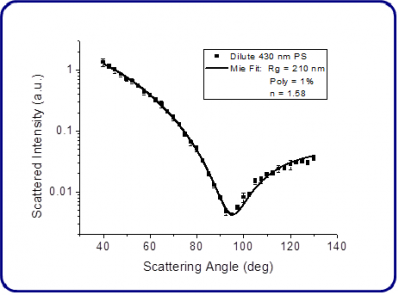
In order to provide a reference for subsequent turbid measurements, SLS data was first gathered for a very dilute suspension of 430 nm polystyrene particles in deionized water (3x10⁻⁷w/w, >99% transmission) over a range of scattering angles. The data shown in Figure 1 were fit using the Mie scattering model (MiePlot, UK) and a visual best-fit with particle refractive index fixed at η = 1.58 gives Rg= 210 nm and 1% polydispersity, in excellent agreement with the nanoparticle product specifications.

Figure 1. SLS data and Mie fit for a dilute sample (3x10⁻⁷w/w, >99% transmission) of 430 nm polystyrene particles in water.
SLS measurements for a turbid sample
With the 3D DLS from LS Instruments, accurate static light scattering measurements in concentrated, highly scattering turbid samples can be performed. However, the undesired contributions from multiply scattered light have to be subtracted from the measured scattered intensity Is. This can be accomplished by performing a combined static and dynamic cross-correlation experiment. In the static measurement, Is is determined as a function of scattering angle q, while the dynamic measurement gives the part of Is which is only singly scattered via the intercept of the cross-correlation function [1]. In the measurement, the reduced intercept
is normalized to
, where
is the intercept measured with a diluted sample without multiple scattering contributions. This automatically corrects for any weak angular dependence of the intercept due to the geometry of the overlap volume and the alignment of the instrument.
The corrected singly scattered intensity as a function of the scattering angle can be calculated as:
(1)
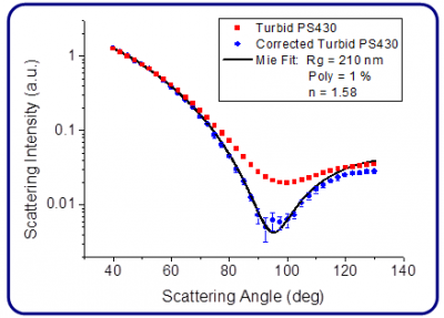
Figure 2 displays the scattered intensity (red squares) measured for a sample of the same 430 nm polystyrene particles but at a significantly increased concentration (5x10⁻⁵w/w), rendering the sample highly turbid (46% transmission). The high degree of multiple scattering for this sample significantly washes out the form factor minimum, and without correction would lead to dramatic errors in estimating sample polydispersity (of nearly an order of magnitude) and mean size. However, by using DLS cross-correlation measurements taken in conjunction with the SLS data and applying Eq. 1, we accurately recover the true particle form factor (blue circles) by extracting only the single-scattering information. This technique becomes even more important when finer features become hidden by multiple scattering that for example, reveal interaction potentials in the sample.

Figure 2. Corrected and uncorrected SLS data along with a Mie fit for a turbid sample of 430 nm polystyrene particles in water (5x10⁻⁵w/w, 46% transmission).
References
[1] C. Urban and P. Schurtenberger, J. Colloid Interface Sci. 207, 150 (1998)
